
Why Keloids Come Back After Surgery and How to Reduce the Risk
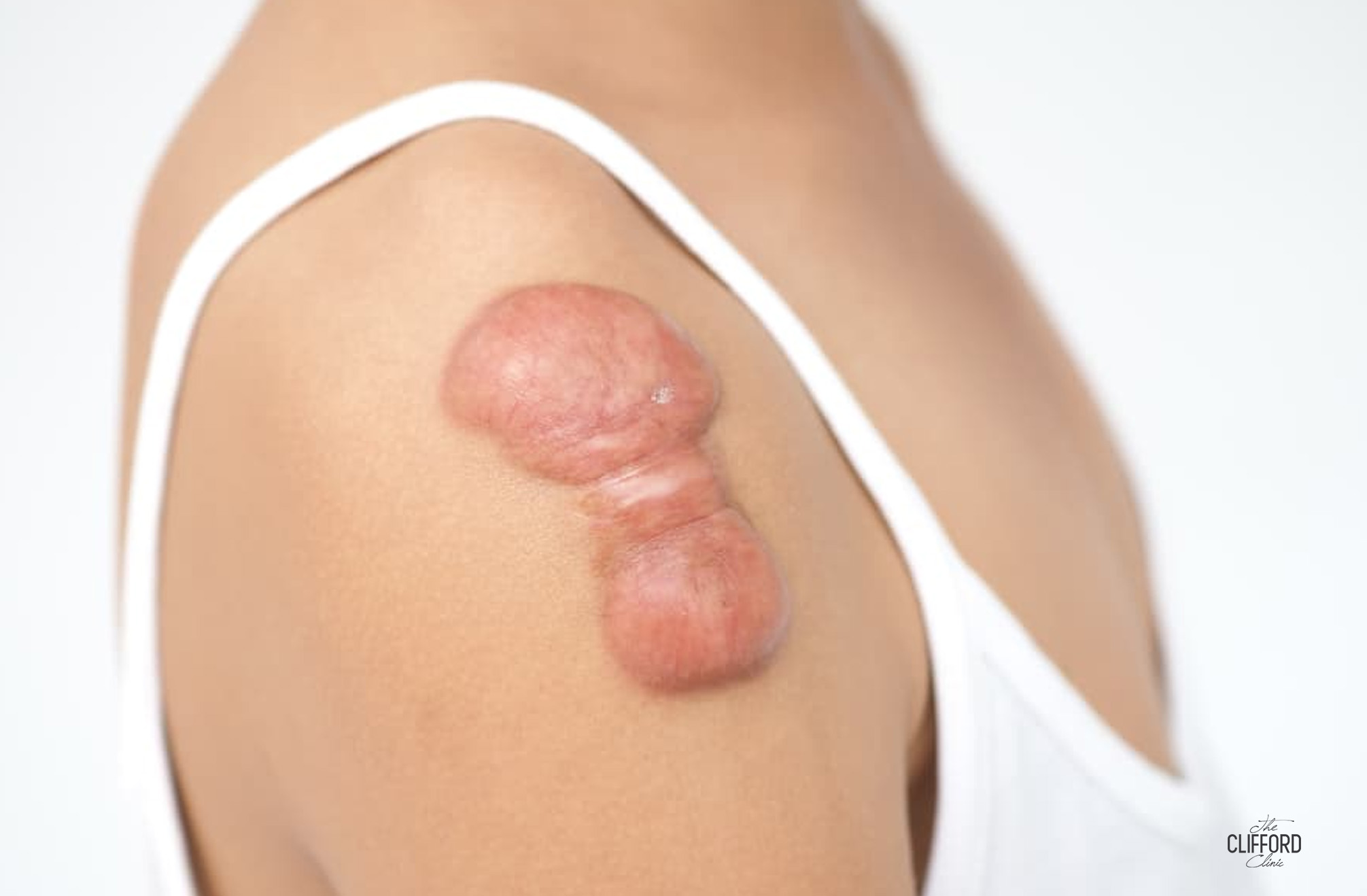
Excision of a keloid is intuitively appealing. The lesion presents as unwanted tissue, and the obvious remedy is to remove it. That intuition is the source of one of the more damaging errors in scar management. Keloids treated by excision alone recur in the great majority of cases, and the recurrent lesion is sometimes larger than the one removed [2][10].
Surgery nevertheless remains an important option, and for large or treatment-resistant keloids sometimes the best one. What separates a durable result from a larger keloid is whether the excision is combined with a planned recurrence-prevention step. This article sets out why excision alone fails, which adjuvant therapies reduce recurrence, and when surgery is appropriate, within our wider keloid scar treatment Singapore resource.
Why Keloid Surgery Alone Almost Always Fails
A keloid is not a foreign body lodged in the skin. It is a product of the way that skin heals. Keloid-prone skin responds to wounding with sustained overproduction of collagen, driven by excessive transforming growth factor beta (TGF beta) signalling, which keeps the fibroblasts, the cells that lay down scar tissue, dividing and depositing collagen long after the wound has closed [1][2].
Excision removes the existing scar by creating a fresh full-thickness surgical wound, often longer than the original injury, in skin that has already demonstrated a keloidal healing response. The surgical trauma itself re-triggers that same response. Because the underlying biology is unchanged, the same fibroblast overactivity acts on a larger wound and frequently produces a keloid larger than the one excised. No evidence-based guideline endorses standalone excision, and a proposal to excise a keloid without a documented recurrence-prevention plan should be declined.
What Influences Keloid Recurrence Risk
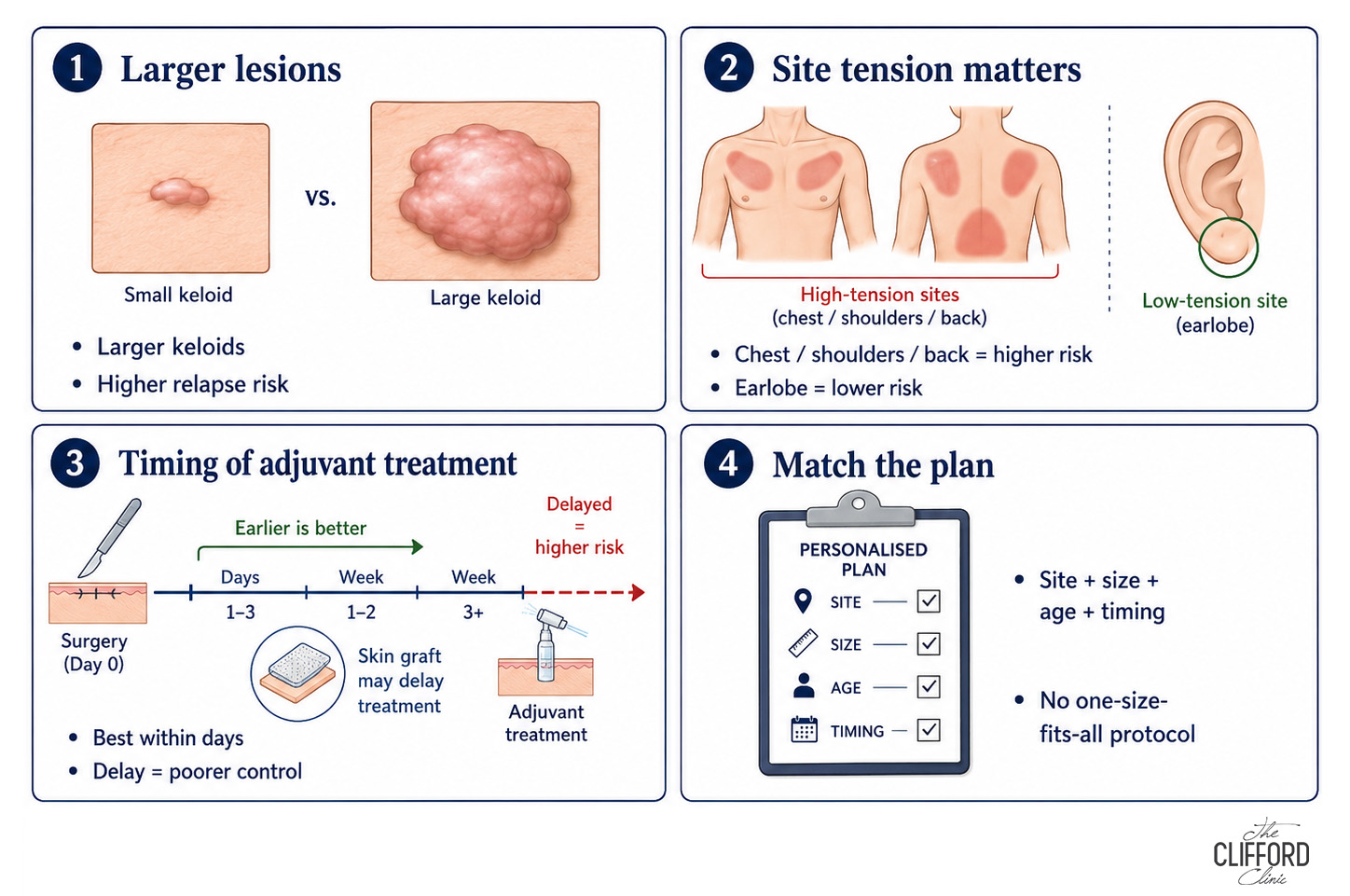
Recurrence risk is not uniform across excised keloids. Larger lesions and those at high-tension sites such as the chest, shoulders and back relapse more readily than small lesions at low-tension sites such as the earlobe. The interval between surgery and adjuvant treatment is a further determinant, because the proliferative burst of fibroblast activity begins within days of wounding, and the adjuvant step is most effective when it interrupts that phase rather than an established scar. Where the defect requires a skin graft the adjuvant schedule is deferred until the graft has taken, which lengthens the unprotected interval. In a series of 834 keloids, larger lesion size, younger age and a longer interval between operation and irradiation were each independently associated with a poorer outcome [12]. The plan is therefore matched to the individual keloid rather than applied as a single protocol [10][11].
Adjuvant Therapy, How Keloid Recurrence Is Reduced
In contemporary practice excision is the first step of a protocol, with the adjuvant therapy selected and scheduled before the operation.
Surgery Plus Immediate Radiotherapy
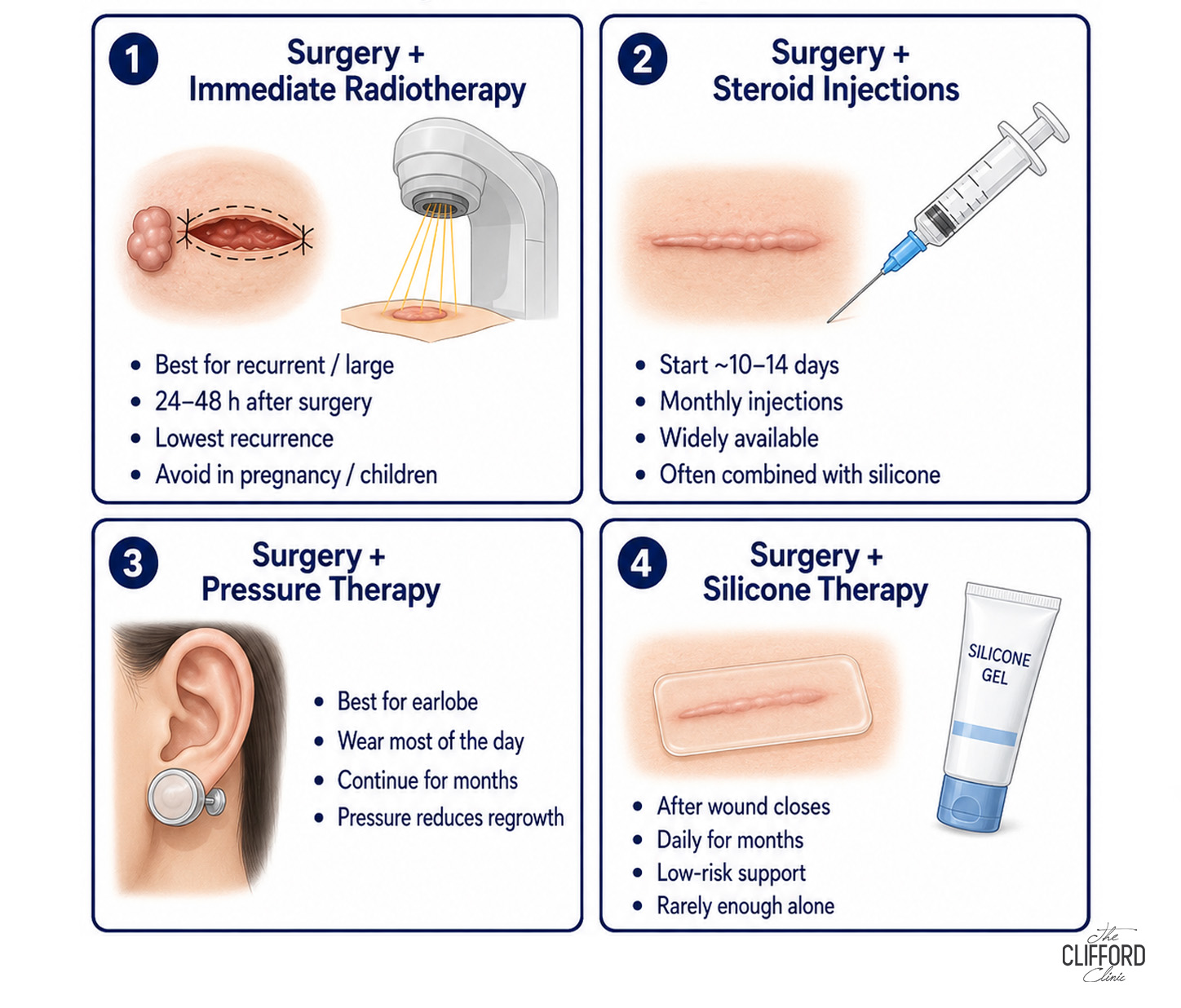
For recurrent, large or treatment-resistant keloids, excision followed by immediate low-dose radiotherapy is among the most effective specialist pathways. Radiation acts on the rapidly dividing fibroblasts in the fresh wound, arresting the cell cycle and triggering apoptosis, the controlled cell death that removes them, so the collagen-producing population is depleted before it can rebuild the keloid. The timing of the first fraction governs the result. In the largest published series radiotherapy was delivered 24 to 48 hours after excision, and was deferred to between 10 and 15 days only where a skin graft had first to take [12].
In 250 resected keloids treated with adjuvant radiotherapy, overall recurrence was 5.6% at a median follow-up of 40 months, and the effect was dose-dependent, with 1.6% recurrence after 20 Gy in five fractions against 9.6% below that dose [10]. A separate series of 100 keloids on the same 20 Gy schedule reported 84.8% local control at a median of 59 months, with acute skin reaction in 3% and late skin sclerosis in 1% [11]. A series of 834 keloids treated with electron-beam radiotherapy reported 88.25% local control and adverse effects in 9.83%, with no radiation-induced cancers observed, and poorer outcomes where treatment started late or the keloid lay at a high-tension site [12]. Reported skin side effects include peeling, itching and change in skin colour. A long-term malignancy risk remains debated rather than demonstrated, so radiotherapy is used selectively and is avoided in children, adolescents and pregnancy [10][11]. The techniques, including proton beam therapy, are covered in our radiation and proton beam therapy guide.
Surgery Plus Steroid Injections
Intralesional corticosteroid suppresses the collagen overproduction that drives regrowth, and evidence favours starting 10 to 14 days after the operation rather than before or during it, with injections repeated at monthly intervals thereafter [2]. This is the most widely available of the adjuvant combinations and is readily combined with silicone. The technique is set out in our steroid injections guide.
Surgery Plus Pressure Therapy
Sustained mechanical pressure reduces blood flow to the healing scar and suppresses regrowth, and the effect depends on total wear time rather than on the appliance itself, so the device is worn for the greater part of each day and continued for several months. Its use is confined to sites where an appliance can be held in place, and it is therefore the principal adjuvant for earlobe keloids, applied as pressure earrings and covered in our ear keloid removal guide.
Surgery Plus Silicone Therapy
Silicone gel or sheeting is applied once the wound has closed and worn daily for several months, and it acts by hydrating the outer layer of the scar and reducing the signals that drive further collagen deposition, at a low risk of adverse effects. It is seldom sufficient on its own after excision, but it reinforces any combination and is the adjuvant a patient can most readily sustain. The evidence is reviewed in our silicone gel sheets guide.
Protocols are commonly combined, for example excision with steroid injections, silicone and pressure, or excision with radiotherapy and silicone, according to the keloid’s history, size and site.
When Keloid Surgery Is Appropriate
Surgery is indicated in four situations. The first is a keloid too large for intralesional injection alone to give an acceptable result. The second is a pedunculated lesion, most often on the earlobe, where the narrow stalk makes complete excision straightforward. The third is failure of first-line treatment after an adequate course. The fourth is symptomatic disease, in which pain or restricted movement warrants debulking. Excision is usually inappropriate as an initial intervention for a small, recently formed keloid that has never been treated, and such lesions should first receive an adequate trial of steroid-led therapy. Smaller keloids may also respond to non surgical treatment alone.
Recovery and Follow-Up After Keloid Surgery
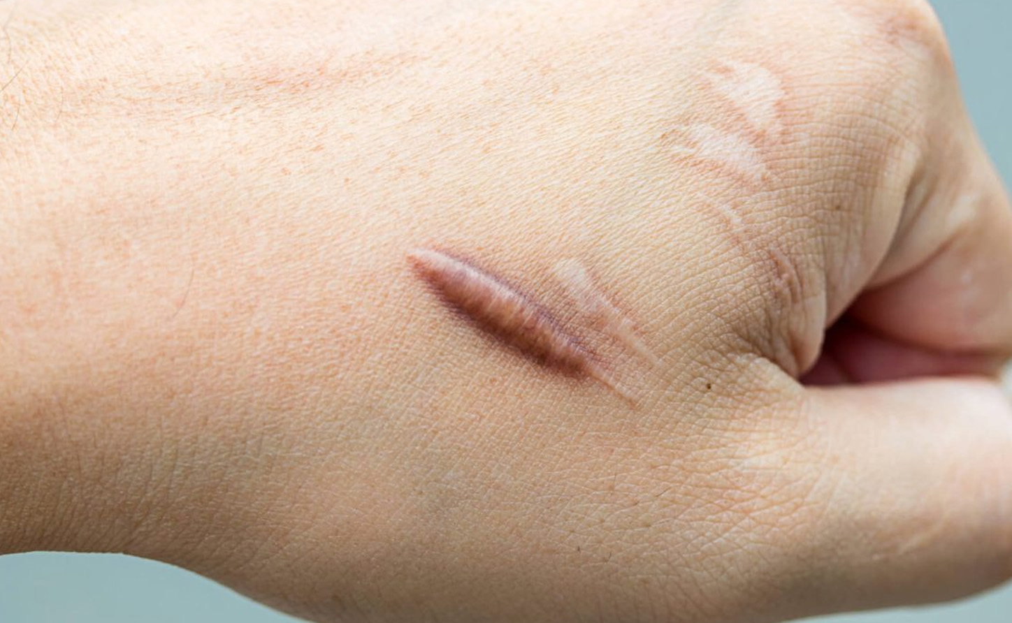
Recovery from the excision itself depends on site and size, and typically involves one to two weeks of basic wound care. Recovery from keloid surgery as a whole is measured in months, however, because the recurrence-prevention phase is the treatment. It comprises scheduled adjuvant sessions, several months of daily silicone or pressure wear, and clinical review continuing for at least a year, because a wound that appears fully healed at an early stage can begin to thicken months afterwards. In a series of 834 keloids, relapse occurred between 6 and 28 months after treatment, at a median of 12 months [12]. Renewed firmness, itching, or a raised edge extending beyond the incision line should be reported between appointments, since early regrowth identified at review is considerably easier to suppress than an established recurrence.
Frequently Asked Questions
What percentage of keloids come back after surgery?
Excision alone recurs in the great majority of cases. With immediate postoperative radiotherapy the published figures are substantially better, with overall recurrence of 5.6% across 250 resected keloids and local control of 84.8% and 88.25% in two larger series [10][11][12]. Combinations with steroids, pressure and silicone also reduce recurrence, although the reported figures vary more widely.
Does keloid removal surgery make it worse?
Excision performed without adjuvant cover can produce a keloid larger than the one removed. Adjuvant therapy is therefore selected and scheduled before the operation rather than considered after it.
How do I stop a keloid from coming back?
Recurrence is reduced by combining excision with an adjuvant therapy, most commonly steroid injections, pressure or silicone, and radiotherapy in difficult cases. The adjuvant is planned before the operation rather than added afterwards, and follow-up is attended so that early regrowth is identified and treated while it remains easy to suppress.
Is radiotherapy after keloid surgery safe?
Low-dose radiotherapy after excision is an established treatment. Acute skin reaction affected 3% and late skin sclerosis 1% in a series of 100 keloids, and a series of 834 keloids recorded adverse effects in 9.83% with no radiation-induced cancers observed [11][12]. A long-term malignancy risk remains debated rather than demonstrated, so the treatment is used selectively and is avoided in children, adolescents and pregnancy.
Can a keloid be removed permanently?
No treatment guarantees permanence, although combination protocols achieve durable control in most patients. The objective is the greatest improvement compatible with the lowest recurrence risk, sustained by continued follow-up.
The Clifford Clinic Approach

Over more than 16 years of treating keloid scars, The Clifford Clinic has found that keloids treated with steroid injections alone almost always recur. The approach used here combines V Beam pulsed dye laser, botulinum toxin and steroid injections, supported by silicone. These address the vascular supply of the keloid, the mechanical tension across the wound and the collagen overproduction at the same time rather than sequentially, and in combination they can prevent recurrence [1][8][9]. Few centres offer V Beam for keloids, and in our experience it is fundamental to preventing their return. It can be delivered safely on darker skin tones, so it remains available across the range of skin types seen in Singapore. Keloids are never removed by surgery alone at this clinic. For large or treatment-resistant keloids, excision combined with radiotherapy is the preferred option. Young keloids are treated early rather than observed, and pigment-safe, skin-tone-aware treatment is selected from the outset. Further information about the clinic is available at cliffordclinic.com and about Dr Bernard Ong at drgerardee.com.
Medically reviewed content. This article is educational and does not replace a consultation. To discuss surgery with adjuvant therapy for a keloid in Singapore, contact The Clifford Clinic.
Related Guides
Keloid scar treatment in Singapore, the complete guide
Steroid injections for keloids
Laser treatment for keloid scars
Ear keloid removal in Singapore
Keloid treatment cost in Singapore
References
1.Laser Therapy of Traumatic and Surgical Scars and an Algorithm for Their Treatment. Lasers in Surgery and Medicine. 2020.
2. Management of Keloids and Hypertrophic Scars. American Family Physician. 2024.
3. Laser Therapy for Treating Hypertrophic and Keloid Scars. The Cochrane Database of Systematic Reviews. 2022.
4. Treatment Response of Keloidal and Hypertrophic Sternotomy Scars, Comparison Among Intralesional Corticosteroid, 5-Fluorouracil, and 585-nm Flashlamp-Pumped Pulsed-Dye Laser Treatments. Archives of Dermatology. 2002.
5. Efficacy and Safety of Glucocorticoid-Based Therapies in the Management of Keloids, A Systematic Review and Meta-Analysis of Clinical Outcomes. Frontiers in Medicine. 2025.
6. Comparing Combination Triamcinolone Acetonide and 5-Fluorouracil With Monotherapy Triamcinolone Acetonide or 5-Fluorouracil in the Treatment of Hypertrophic Scars, A Systematic Review and Meta-Analysis. Plastic and Reconstructive Surgery. 2024.
7. Comparative Efficacy of Intralesional Therapies for Keloid Scars, A Network Meta-Analysis. Annals of Medicine. 2026.
8. Assessment of Intralesional Injection of Botulinum Toxin Type A in Hypertrophic Scars and Keloids, Clinical and Pathological Study. Dermatologic Therapy. 2022.
9. Botulinum Toxin Type A Versus 5-Fluorouracil in Treatment of Keloid. Archives of Dermatological Research. 2021.
10. Dose Effect in Adjuvant Radiation Therapy for the Treatment of Resected Keloids. International Journal of Radiation Oncology, Biology, Physics. 2018. 11. A Retrospective Study of Hypofractionated Radiotherapy for Keloids in 100 Cases. Scientific Reports. 2021. 12. Hypofractionated High-Energy Electron Beam Radiotherapy for Keloids. Journal of Radiation Research. 2015.
Author Bio
Dr Gerard Ee

Dr Gerard Ee is the Medical Director of The Clifford Clinic in Singapore. He earned his medical degree from St George's University of London and built his surgical experience across the Singapore General Hospital, National University Hospital, and Mount Sinai Hospital in New York. He holds Membership of the Royal College of Surgeons (Edinburgh) and a Postgraduate Diploma in Dermatology (Cardiff), and is fully accredited in aesthetic procedures including botulinum toxin, fillers, lasers, and light therapies. A strong advocate of minimally invasive, natural-looking results, Dr Ee has published in several medical and scientific journals and presented his research in cities including Taipei, Las Vegas, and London.
See all posts by Dr Gerard Ee »